
Fig. 5.
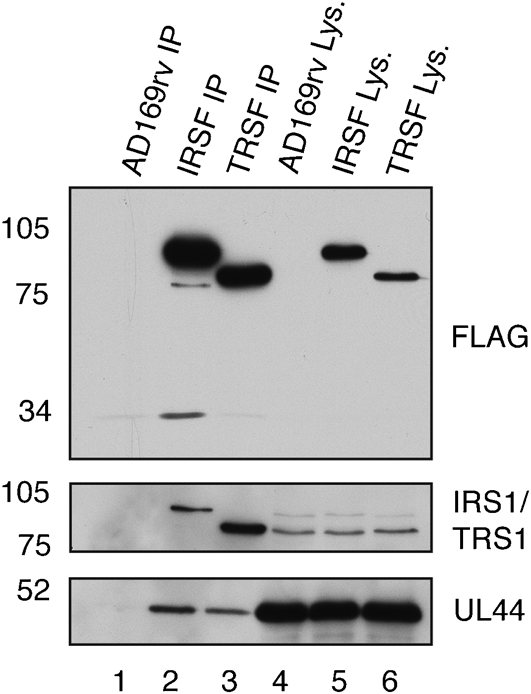
Detection by Western blotting of IRS1 and TRS1 in protein immunoprecipitated from IRSF- and TRSF-infected cell lysate. Lysate from AD169rv-infected (lane 4), IRSF-infected (lane 5) and TRSF-infected (lane 6) cells and protein immunoprecipitated using an anti-FLAG antibody from those lysates (lanes 1–3, respectively) were separated on a 10 % polyacrylamide gel. Proteins in each lane were examined by Western blotting for the presence of FLAG-tagged protein (top panel), IRS1 or TRS1 (middle panel) or UL44 (bottom panel) using antibodies recognizing these proteins, as indicated to the right of the figure. Note that, in the middle panel, a combination of antibodies recognizing IRS1 or TRS1 was utilized. The positions of molecular mass markers (in kDa) are indicated to the left of the figure.