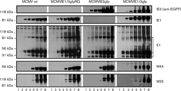
Fig. 2.
Confirmation of the resultant MCMVs with EGFP-fused IE3 gene by WB. NIH3T3 cells were mock-infected (lane 1) or infected with MCMV (indicated on the top) for 3 h (lane 2), 6 h (lane 3), 12 h (lane 4), 24 h (lane 5), 36 h (lane 6), 48 h (lane 7) and 72 h (lane 8). The whole-cell lysis samples were separated by PAGE and transferred to nitrocellulose membranes for WB. Blots were first probed with anti-EGFP to detect ∼108 kDa IE3–EGFP (top), then stripped and reprobed with antibodies against the proteins indicated on the right. MCMVIE3gfp did not produce IE1 protein. Note that the revertant (MCMVIE1/3gfpRQ) produced IE1, but did not produce IE3–EGFP. Also note that the presence of IE1 protein appeared to delay the production of IE3–EGFP (top right panel), which in turn may have delayed the production of M25 protein (bottom right panel).

