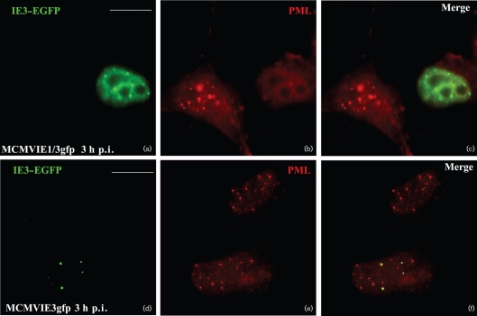
Fig. 3.
EGFP-fused IE3 gene products and ND10 after viral infection. NIH3T3 cells were infected with MCMVIE3gfp (a–c) and MCMVIE1/3gfp (d–f) for 3 h, fixed and permeabilized, and stained with anti-PML antibody to detect ND10, followed by Texas Red-labelled secondary antibody. The results show PML alone (red; b and e), IE3–GFP alone (green; a and d) and the merged images (c and f). Bars, 10 μm.

