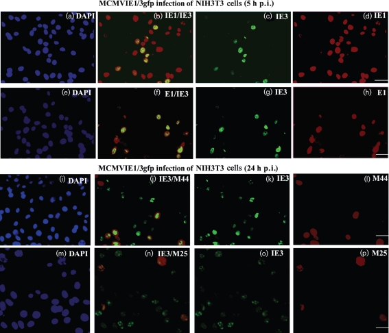
Fig. 4.
Immunofluorescent assay for detection of viral proteins after infection. NIH3T3 cells on coverslips were infected with MCMVIE1/3gfp for 5 h (a–h) or 24 h (i–p) and fixed. DAPI staining (panels a, e, i and m) was used to show the total number of cells (infected and uninfected). IE3 was detected as green fluorescence due to the fused EGFP (c, g, k, and o). Other viral proteins (IE1 and E1, early proteins; M44 and M25, early-late proteins) were stained in red (d, h, l and p). The merged images are shown in (b), (f), (j) and (n). Bars, 20 μm.

