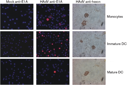
Fig. 1.
Expression of HAdV E and L antigens in infected cells (strain BB2000-61). Expression of E and L antigens was visualized 3 and 5 days post-infection. Cells were either mock infected or infected with BB2000-61. Mock is supernatant from cells treated exactly as infected ones except for the addition of HAdV. The m.o.i. of 1 resulted in 31 %, m.o.i. of 10 in 39 %, m.o.i. of 20 in 29 % and m.o.i. of 50 in 31 % positive-stained immature DC with E1A-staining. E1A antibody was diluted 1 : 100. Hexon specific antibody was diluted 1 : 200. Secondary antibody, anti-mouse immunoperoxidase was diluted 1 : 500. Counterstaining with DAPI.

