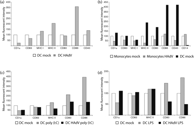
Fig. 2.
Expression of immunologically relevant surface markers after HAdV infection with an m.o.i. of 10. (a) Freshly isolated DC were stained with antibodies specific for the designated markers or differentiated into mdDC by the addition of IL-4 and GM-CSF and cultivation for 7 days, or infected with HAdV and cultivated for a further 7 days in the presence of IL-4 and GM-CSF then stained and analysed by flow cytometry. (b) Freshly isolated monocytes were infected with the strain BB2000-61 and cultivated for a further 5 days. (c, d) Mature DC were infected with the same virus. The experiments were performed twice.

