Figure 2.
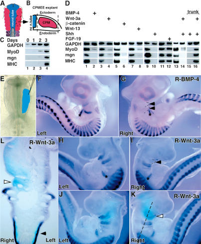
Ectopic BMP, Wnt, and Hedgehog signals can block myogenesis in the cranial paraxial mesoderm. (A) Diagram of a stage 10 chick embryo, showing the location of the cranial paraxial mesoderm (red) adjacent to the neural tube (blue) and anterior to the first pair of somites (purple). (B) Scheme of the germ layers included in the dissected tissue, showing the cranial paraxial mesoderm covered dorsally by the ectoderm and ventrally by the endoderm, termed CPMEE. (C) Skeletal myogenesis in CPMEE explants cultured in vitro. CPMEE explants were dissected and cultured in vitro for the indicated number of days prior to harvest. RT-PCR analysis was performed to monitor expression of the indicated genes. (D) The effect of various signaling molecules on myogenesis in cranial and trunk paraxial mesoderm. Explants of either CPMEE (lanes 1-14) or somites I-III (lanes 15,16) isolated from stage 10 chick embryos were cultured in vitro. Explants were cultured in medium containing either BMP-4 (lane 2), Shh (lanes 10,14,15,16), or FGF-19 (lane 12), or infected with a nondefective retrovirus (RCAS) encoding either GFP (lanes 3,5,7,13,15), Wnt-3a (lanes 4,14,16), a stabilized form of β-catenin (lane 6), or Wnt-13 (lane 8). A representative experiment is shown; however, similar results were obtained in at least four independent experiments. (E) Cranial paraxial mesoderm of stage 9 embryos targeted for infection by RCAS viruses (blue) in F-L. (F,G) Effect of RCAS-BMP-4 infection of the cranial paraxial mesoderm. MyoD expression in either the control side (left) or the RCAS-BMP-4-infected side (right), respectively. The asterisk marks developing tongue muscles that migrate from occipital somites, closed arrowheads identify BMP-4-infected branchial arches. (H,I) Effect of RCAS-Wnt-3a infection of the cranial paraxial mesoderm. Myf-5 expression in either the control side (left) or the RCAS-Wnt-3a-infected side (right), respectively. The asterisk marks developing tongue muscles that migrate from occipital somites. The closed arrowhead identifies Wnt-3a-infected branchial arch. (J,K) Secondary in situ hybridization to detect ectopic viral gene expression (light blue; open arrowhead). Light blue staining in the branchial arch region of J is from staining on the contra-lateral RCAS-Wnt-3a-infected right side of the embryo; see L. (L) A frontal section of the embryo depicted in J and K (at the level labeled with a dashed line) documents viral expression (light blue) in both the right side of the branchial arches (which lack Myf-5 expression; open arrowhead) as well as the somitic mesoderm (which retains Myf-5 expression; closed arrowhead).

