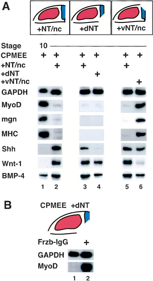
Figure 3.
Inhibition of myogenesis in the cranial paraxial mesoderm by signals from the dorsal neural tube. (A) Diagrams of transverse sections through explants show the cranial paraxial mesoderm (red) in relation to the adjacent axial tissues (blue). RT-PCR analysis of gene expression in explants that had been isolated from stage 10 chick embryos and cultured for 4 d in either the absence (lane 1) or the presence of the adjacent neural tube and notochord (NT/nc; lanes 2,3,5), the dorsal neural tube (dNT; lane 4) or the ventral neural tube and notochord (vNT/nc; lane 6). Skeletal myogenesis was observed in 1/15 explants cocultured with NT/nc (lanes 2,3,5), in 0/8 explants cocultured with dNT (lane 4), and in 5/5 explants cocultured with vNT/nc (lane 6). (B) Administration of Frzb-IgG can induce MyoD expression in CPMEE explants cultured in the presence of the dNT. CPMEE plus the adjacent dorsal neural tube were isolated from stage 10 chick embryos and were cultured in either the absence (lane 1) or presence (lane 2) of Frzb-IgG. MyoD induction was observed in 40% of such explants (n = 10).

