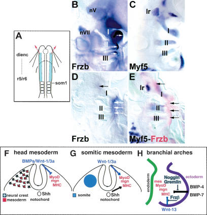
Figure 7.
Myogenic bHLH genes are expressed in the branchial arches after ablation of the cranial neural crest; a model for cranial skeletal muscle induction. (A) Scheme of in ovo neural crest cell ablation. At stage 8, the dorsal half of the neural plate from diencephalic levels to rhombomere 5/6, anterior to the first somite (som1), was excised bilaterally as indicated. (B-E) Lateral views of the BA region of either control (B,C) or operated (D,E) embryos at stage 18-19 (dorsal is to the left, anterior to the top). (B) Control embryo stained for Frzb. Note the widespread expression of Frzb, including the trigeminal (nV) and facial (nVII) ganglia, the neural crest cells in the BA mesenchyme, and the ectoderm covering the trigeminal and the distal region of the BAs (arrows). (C) Expression of Myf-5 in a control embryo. The anlagen of the muscles in BA I-III and of the lateral rectus muscle (lr) are indicated. (D) Neural crest ablated embryo stained for Frzb. Although neural crest cell-associated expression of Frzb is significantly diminished in the head region, Frzb is still present in the ectoderm overlying the trigeminal ganglia and the distal ectoderm of the BAs (arrows). (E) Neural crest ablated embryo stained for Myf-5 in blue and Frzb in red. Note that Myf-5 expression is still detectable both in the lateral rectus and in the BAs. In this embryo, low levels of Frzb are detectable throughout the BAs and higher levels are found in the ectoderm overlying the trigeminal ganglia and the distal ectoderm of the BAs (arrows). (dienc) diencephalon; (r5/6) rhombomeres 5 and 6. (F-H) Head muscle formation is cued by a combination of negative and positive signals from surrounding tissues. (F) Cranial neural crest cells (CNC, shown as grey) migrate away from the dorsal neural tube and into the region of the cranial paraxial mesoderm (red). While lying adjacent to the neural tube, muscle formation in the cranial paraxial mesoderm is blocked by BMP and Wnt signals from the dorsal neural tube (G) In the trunk, Wnt signals from the dorsal neural tube in combination with Shh signals from the notochord and floor plate promote epaxial myogenesis in the somites. (H) At sites of head muscle formation such as the BAs, CNC cells, which have migrated from the dorsal neural tube, surround a core of muscle precursors derived from the cranial paraxial mesoderm. BMP-4, BMP-7, and Wnt-13 signals from the BA ectoderm repress skeletal muscle differentiation in the mesodermal core. The BMP antagonists Noggin and Gremlin work together with the Wnt antagonist Frzb, which is expressed by both CNC cells and other BA tissues, to override BMP and Wnt inhibition and thereby promote skeletal myogenesis.

