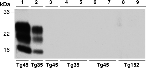
Fig. 2.
Failure to detect PrPSc in the brain of CWD prion-inoculated transgenic mice. The high sensitivity immunoblot using anti-PrP monoclonal antibody 3F4 shows PK-digested sodium phosphotungstic acid pellets recovered from 10 % (w/v) transgenic mouse brain homogenates. Lanes 1 and 2, positive controls showing efficient recovery of PrPSc after spiking 2 μl 10 % (w/v) BSE-inoculated 129MM Tg45 and 129MM Tg35 transgenic mouse brain homogenates (Asante et al., 2002) into 100 μl 10 % (w/v) uninfected 129MM Tg45 and 129MM Tg35 mouse brain homogenates, respectively. Lane 3, PK-digested sodium phosphotungstic acid pellet from 250 μl 10 % (w/v) brain homogenate from a 129MM Tg45 mouse inoculated with normal mule deer brain. Lanes 4–9, PK-digested sodium phosphotungstic acid pellets from 250 μl 10 % (w/v) brain homogenates from 129MM Tg35, 129MM Tg45 and 129VV Tg152 mice inoculated with CWD-infected mule deer brain.

