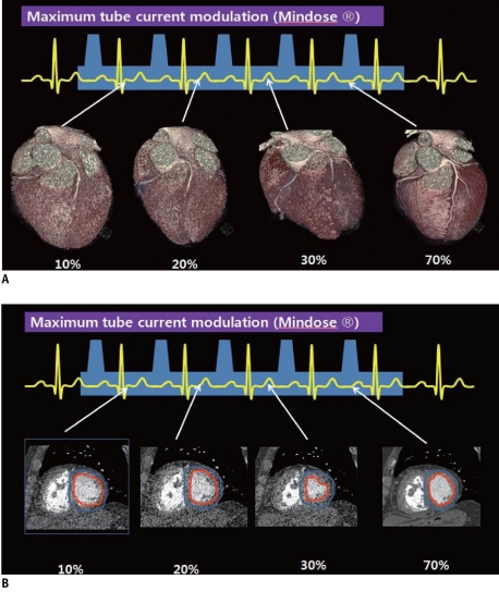
Fig. 4.
Difference of image quality depending on exposed maxinum tube current.
A. Coronary artery anatomy quality was good at 100% of maximum tube current during mid-diastole and poor outside this phase with 4% of maximum tube current. B. Differentiating endocardium from lumen is available during all cardiac phase including phase with 4% of maximum tube current

