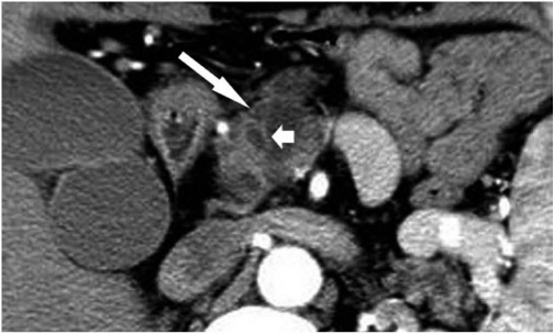
Fig. 1.
Transverse CT scan obtained in 62-year-old man with pancreatic ductal adenocarcinoma with cystic features.
Image was obtained after intravenous injection of contrast material demonstrated irregular multicystic lesion (long arrow) in head of pancreas. Wall is thick and enhancing on this contrast-enhanced image. Note septum (short arrow).

