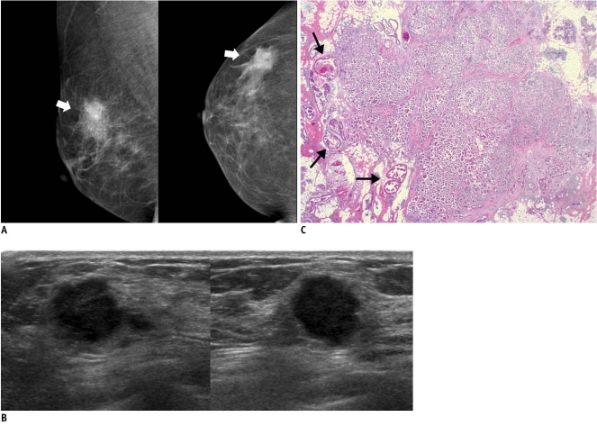
Fig. 1.
46-year-old woman with palpable mass in her right breast.
A. On mammography (left: mediolateral oblique view, right: craniocaudal view), irregular ill-defined hyperdense mass (arrows) is seen on area corresponding to palpable abnormality. B. On sonography (left: transverse view, right: longitudinal view), mass is determined to be round microlobulated hypoechoic mass with nonparallel orientation and classified as BI-RADS category 4c. Result of sonography-guided 14-gauge core needle biopsy are consistent with invasive ductal carcinoma, which is considered as concordant malignancy. C. Histologic features on low power view indicate invasive ductal carcinoma with peripheral intraductal component (arrows) without microcalcification. Surrounding breast parenchyma shows fatty changes (Hematoxylin & Eosin staining; original magnification, × 10).

