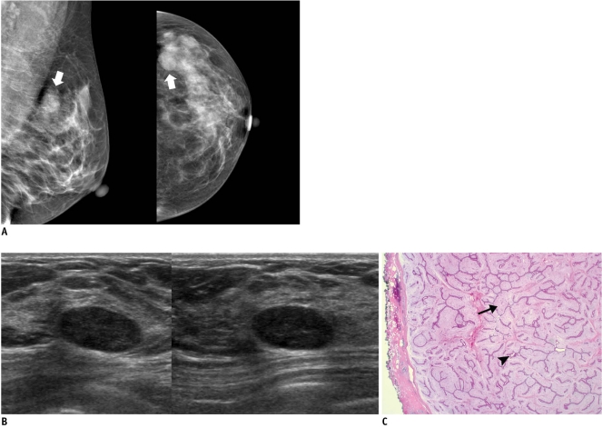
Fig. 3.
45-year-old woman with mass in her left breast.
A. On mammography (left: mediolateral oblique view, right: craniocaudal view), oval circumscribed mass (arrows) is seen in upper outer portion of her left breast. B. On sonography (left: transverse view, right: longitudinal view), oval circumscribed hypoechoic mass is seen and classified as BI-RADS category 3. Sonography-guided 14-gauge core needle biopsy was performed at request of patient and pathologic result indicated fibroadenoma, which is considered to be concordant benign lesion. However, in lieu of follow-up image, surgical excision was performed. C. Photomicrograph of microscopic specimen after surgical excision shows sharply defined border with both glandular (arrowhead) and stromal proliferation (arrow), mainly showing intracanalicular growth pattern. Final diagnosis was fibroadenoma (Hematoxylin & Eosin staining; original magnification, × 10).

