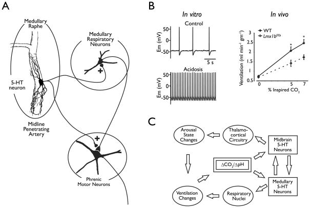
Figure 5.
Central 5-HT neurons are chemoreceptors. A) Push-pull model of the role of chemosensitive medullary raphe neurons in respiratory chemoreception. It is proposed that the two subtypes of chemosensitive raphe neurons act in opposite ways to influence respiratory output within the medulla and in phrenic motor neurons. The brainstem sites are not specified in this model, but may include the nucleus tractus solitarius, nucleus ambiguus, pre-Botzinger complex and hypoglossal motor nucleus. Reproduced with permission from Richerson et al (2001). B) Left: Membrane potential under control conditions (5% CO2, pH 7.4) and during hypercapnic acidosis. Reproduced from Wang et al, (2002). Right: Ventilation during normoxic hypercapnia conditions (n = 10 WT, 8 Lmx1bf/f/p). Modified from Hodges et al (2008). C) Schematic depicting the proposed model by which serotonergic neurons mediate ventilatory and vigilance state changes in response to changes in CO2/pH (ΔCO2/ΔpH). Increased CO2 (due to rebreathing, airway obstruction or apnea) leads to decreased pH and activation of midbrain serotonergic neurons. These neurons in turn activate thalamocortical circuitry resulting in an elevation of arousal state. Increased CO2 also leads to activation of medullary serotonergic neurons, which stimulate respiratory nuclei to increase ventilation. Both of these pathways serve to restore CO2 homeostasis. 5-HT, serotonin. Reproduced with permission from Buchanan et al (2008).

