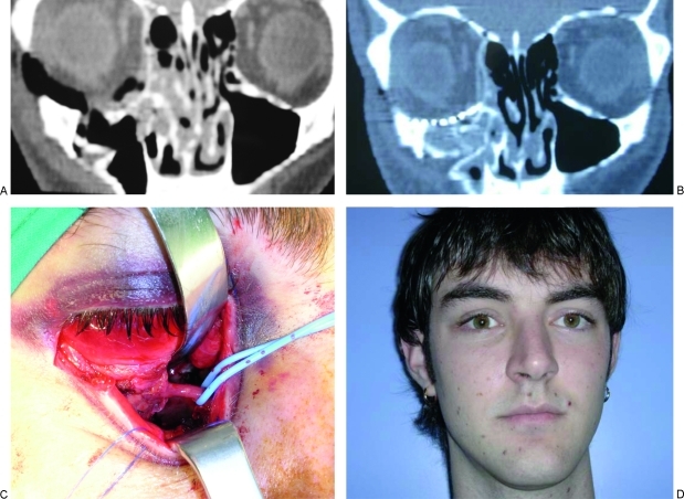
Figure 3.
Case 2. (A) Preoperative CT scan of a 21-year-old man with fracture of the medial wall and floor of the orbit. (B) Postoperative CT scan showing the reconstruction of the floor of the orbit with titanium mesh and the medial wall with PDS sheets. (C) Retroseptal inferior fornix and transcaruncular dissection without detachment of the inferior oblique muscle. (D) Six-month postoperative view of the patient.

