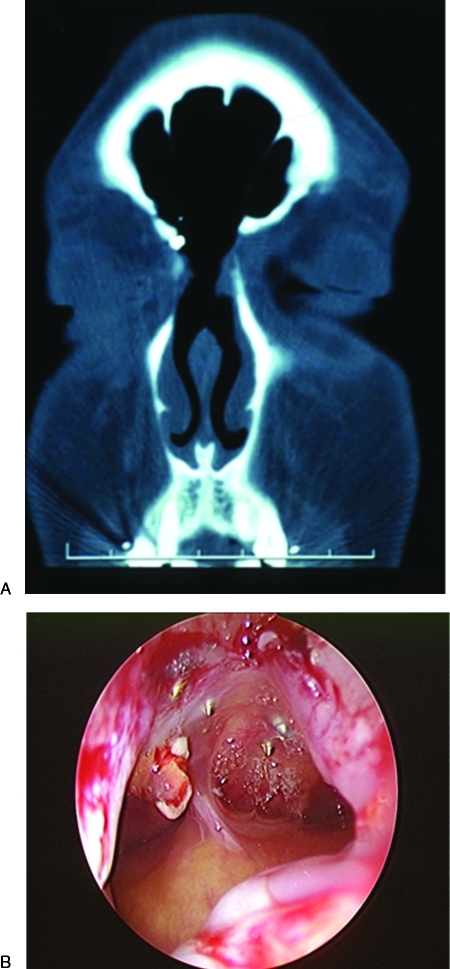
Figure 6.
(A) Postoperative coronal computed tomography scan after unilateral extended frontal sinusotomy (Draf type III) demonstrating frontal sinus ventilation. (B) Endoscopic examination in the office at 6 months demonstrating patency of the open reduction and internal fixation. The screws used for fixation of the anterior table fracture are seen penetrating the wall of the frontal sinus.

