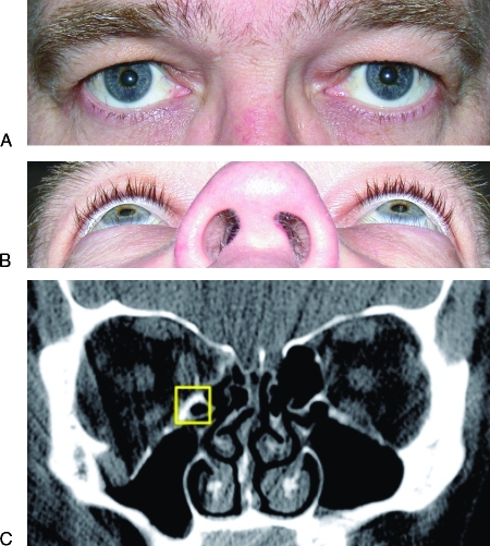
Figure 5.
(A) Close-up frontal view of patient's eyes showing no signs of posttraumatic enophthalmos 6 months following the injury despite having a large right-sided orbital floor and medial wall fracture (Fig. 5C). The patient had no surgery. (B) Close-up worm's-eye view of same patient as seen in Fig. 5A showing no evidence of posttraumatic enophthalmos following large orbital fracture on right side. The patient had no surgery. (C) Coronal computed tomography scan of same patient as seen in Figs. 5A and 5B performed acutely after right-sided orbital floor and medial wall fractures. Total fracture area measures 6.30 cm2. There is minimal rounding seen in the inferior rectus muscle possibly due to either minimal soft tissue herniation or an intact medial orbital buttress (uncinate process of maxilla), which is outlined in the yellow square. Despite having a large fracture, this patient did not develop posttraumatic enophthalmos.

