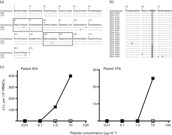
Fig. 3.
Sequence mutants in targeted epitopes. (a) An alignment of the core region is shown. The upper line indicates the group consensus. The lower lines indicate donors 304 and 379 with mutations within targeted epitopes indicated. Dots indicate amino acids identical to the consensus sequence. (b) An alignment of the core region for cloned donor 304 is shown. Each clone was compared with the bulk sequencing product. The frequency of the variant within the epitope 61–80 is indicated by shading: A68V was observed in the majority of the sequenced population. (c) Peptide titrations using PBMCs from donors 304 and 379, using wild-type (▪) and mutant (□) peptide as indicated in Fig. 3(a). The assays were performed as in Fig. 1.

