
Figure 1.
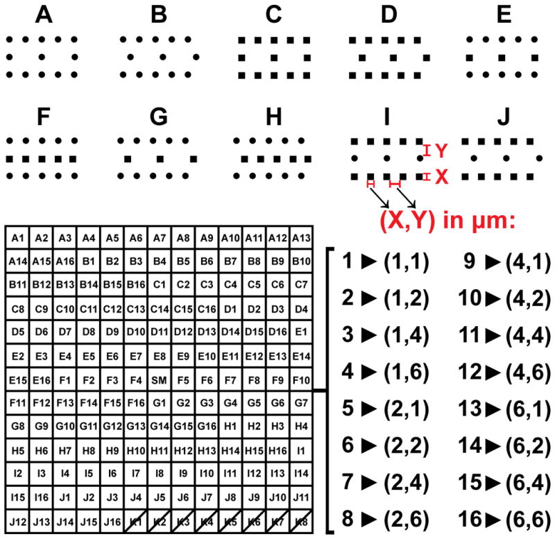
Topographical library details: 10 pit morphologies (A–J) were replicated with 16 different combinations of size (X, 1–6 μm) and spacing (Y, 1–6 μm), giving 160 unique patterned PDMS substrates for cell growth, each with a uniform pit depth of 2.4 μm. Smooth regions were present in the center of the array (SM), and in the regions between patterns. Pattern K was excluded from all analyses due to poor cell attachment.