
Figure 4.
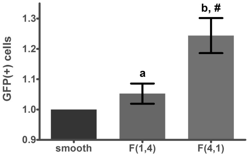
Flow cytometry corroborates the results presented in Figure 3a; F(4,1), a pattern with small interfeature spacing (1 μm), supported 25% higher transfection efficiency than smooth PDMS and F(1,4), a substrate with large interfeature spacing (4 μm).