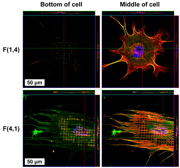
Figure 6.
Confocal microscopy revealed NHDFs exploring the bottom of 1 and 4 μm diameter pits. The appearance of “holes” in the cell membrane of cells cultured on pattern F(4,1) is a consequence of the apical and basal cell surfaces bending below the focal plane, whereas cells on F(1,4) only reached the bottom of pits with their basal membrane.

