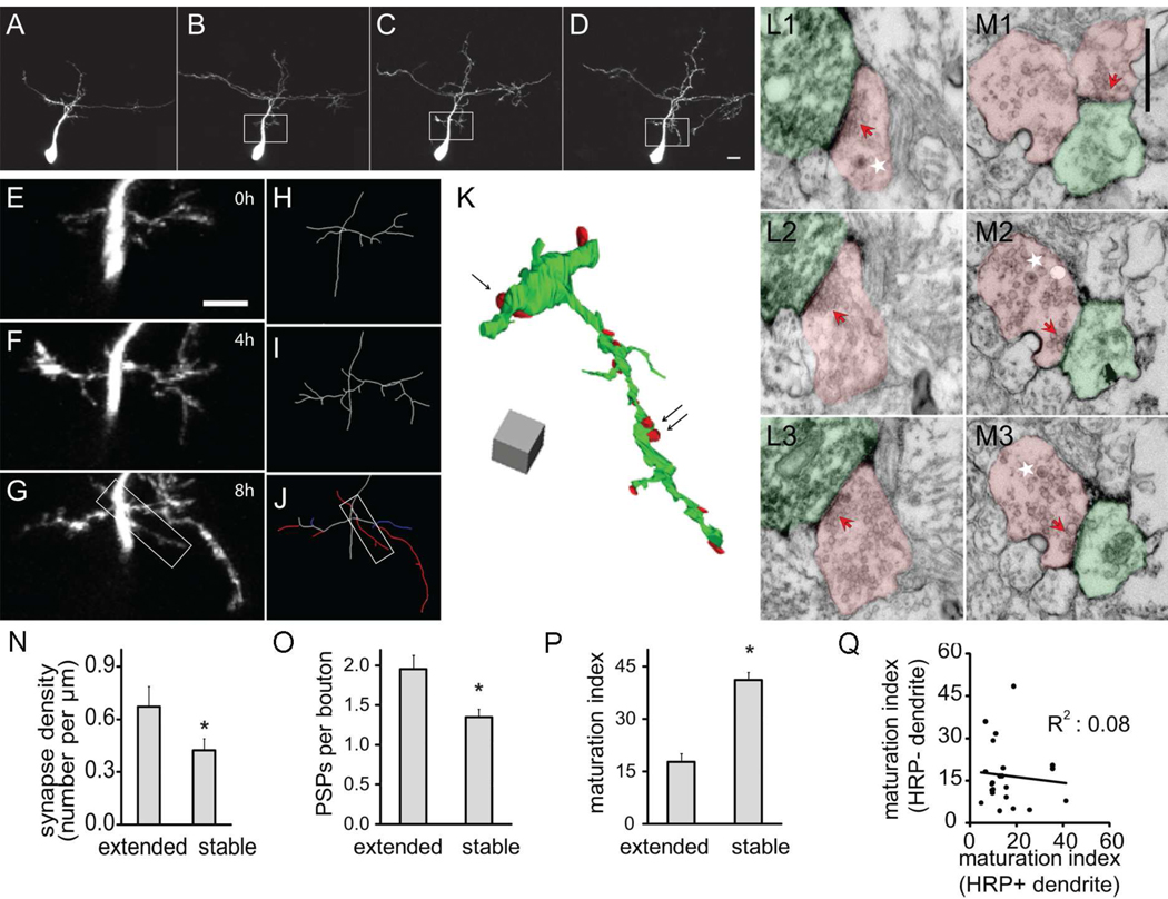
Figure 5. Rapid synapse formation on extended dendritic branches.
(A–D) Two-photon time-lapse images of an optic tectal neuron from a stage 47 tadpole collected on the second day (A) and 0h, 4h, and 8h of the third day (B–D) after electroporation. (E–G) High magnification view of the region in white boxes of B–D. (H–J) Drawings of the partial dendritic arbor of the neuron shown in E–G. Branches are color-coded according to their dynamics over the previous 4h in J. white: stable; red: extended; and blue: retracted. (K) 3D reconstruction of the dendritic arbor shown in the white box of J. (L–M) Serial EM sections through axon terminals contacting branches that were stable (L1–L3) or extended (M1–M3) over the last 4h. The locations of these two synapses are marked by the single arrow and double black arrows, respectively, in K. Axons and dendrites are marked in red and green. Dense core vesicles are marked by white stars and synaptic sites are marked by red arrows. Scale bar in M1 is 500nm and applies to L1–M3. (N) Number of postsynaptic partners of axonal boutons contacting stable and extended branches. (O) The number of postsynaptic profiles (PSPs) per axonal bouton contacting stable and extended dendritic branches. (P) Maturation index of synapses on stable and extended dendritic branches. (Q) Paired comparison of maturation index of divergent synapses from the same axonal bouton contacting mHRP-positive and negative dendrites. mHRP positive dendrites extended within past 4 hours.

