
Figure 2.
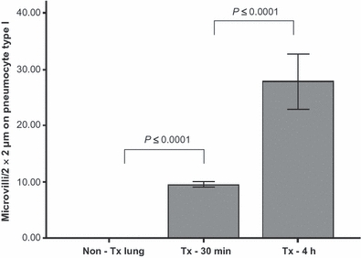
Number of microvilli (MV) per 2 × 2 μm on pneumocytes type I at different time points: non-Tx lung calculated as time zero, Tx-30 min, Tx-4 h after reperfusion. Tx lungs 4 h after reperfusion show significant higher numbers of MV compared to Tx lungs 30 min after reperfusion (27.9 ± 4.9 vs. 9.5 ± 0.5; P ≤ 0.0001), and Tx lung 30 min after reperfusion showed significantly more numbers of MV compared to non-transplanted lung (9.5 ± 0.5 vs. 0.0 ± 0.0; P ≤ 0.0001). Non-Tx Lung, non-transplanted lung; Tx-30 min, transplanted lung, 30 min after reperfusion; Tx-4 h, transplanted lung, 4 h after reperfusion.