
Figure 4.
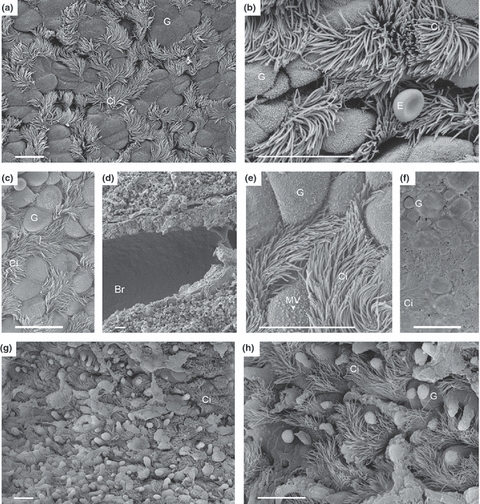
Respiratory tract from right, non-transplanted lung, and transplanted lung, 30 min and 4 h after reperfusion. (a, b) right, non-transplanted lung, normal bronchial tissue with goblet cells and cilia-cells, an erythrocyte (E) as a reference cell lying over bronchial tissue (b); (c–f) transplanted graft 30 min after reperfusion; Microvilli (MV) at the surface of goblet cells have a rounder form and are shorter and clumped (e, arrow) compared to MV depicted in b. The epithelium is partially covered with fluid collection because of oedema (f). (g, h) transplanted graft 4 h after reperfusion shows bronchioles with increased exocytosis of goblet cells, and a decline in numbers of cilia-cells. Br, bronchus; G, goblet cell; Ci, ciliated cell; E, erythrocyte. (a–h) Scale bar = 10 μm.