
Figure 6.
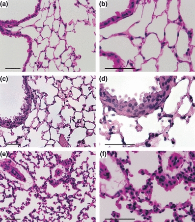
Histology, haematoxylin and eosin (H&E) of corresponding samples from SEM: (a, b) right, non-transplanted lung with normal histoarchitecture. (c, d) Thirty minutes after reperfusion with few infiltrates of mononuclear cells and moderate thickening of the alveolar wall. (e, f) Four hours after reperfusion with increased accumulation of leucocytes and macrophages, paralleled by perivascular oedema and more severe thickening of the alveolar wall. (a–f) Scale bar = 50 μm.