
Figure 7.
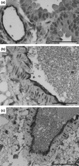
Immunohistochemical staining of ICAM-1. Non-transplanted lungs show a fine lining of ICAM-expression on the endothelium of the vessel. In contrast, transplanted lungs that were exposed to 2 h of ischaemia and 30 min of reperfusion, the endothelium increasingly stains positive for ICAM-1 (b) while transplanted grafts at 4 h of reperfusion revealed a strong staining for ICAM-1 (c). One representative section for each of two grafts is shown. (a–c) Scale bar = 50 μm.