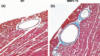
Figure 1.

Masson trichrome stain of mid-ventricular coronary artery cross sections from wild-type (WT) and MMP-2 transgenic (MMP2 TG) mice at 8 months. (a) Cross section of WT coronary artery showing normal diameter, minimal branching and normal amounts of perivascular fibrosis. (b) Cross section of MMP-2 transgenic showing coronary artery dilatation, multiple primary and secondary branching arteries and large amounts of perivascular fibrosis (×200).
