
Figure 4.
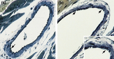
Verhoeff’s elastin stain of mid-ventricular coronary artery cross sections at 8 months of age. (a) WT control with intact internal elastic lamina (arrow). (b) MMP-2 TG: The internal elastic lamina is fragmented and disrupted (arrows). (a, b × 300, inset ×500).