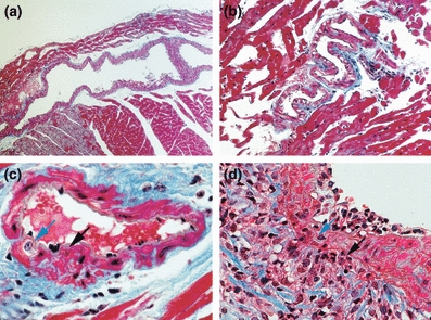
Figure 5.

Characterization of coronary artery ectasia in MMP-2 TG mice. (a) Longitudinal section of a mid-ventricular coronary artery from an 8-month-old transgenic mice, demonstrating massive, localized arterial dilatation with ectasia. (b) Severely ectatic coronary artery with perivascular cellular infiltrates. (c) Focus of medial disruption (black arrow) immediately adjacent to proliferating vascular smooth muscle cell (blue arrow). (d) Focus of profound perivascular inflammatory cell infiltrate (black arrow) extending through the vascular media layer (blue arrow). (Masson trichrome stain; a × 50; b × 100; c, d × 400).
