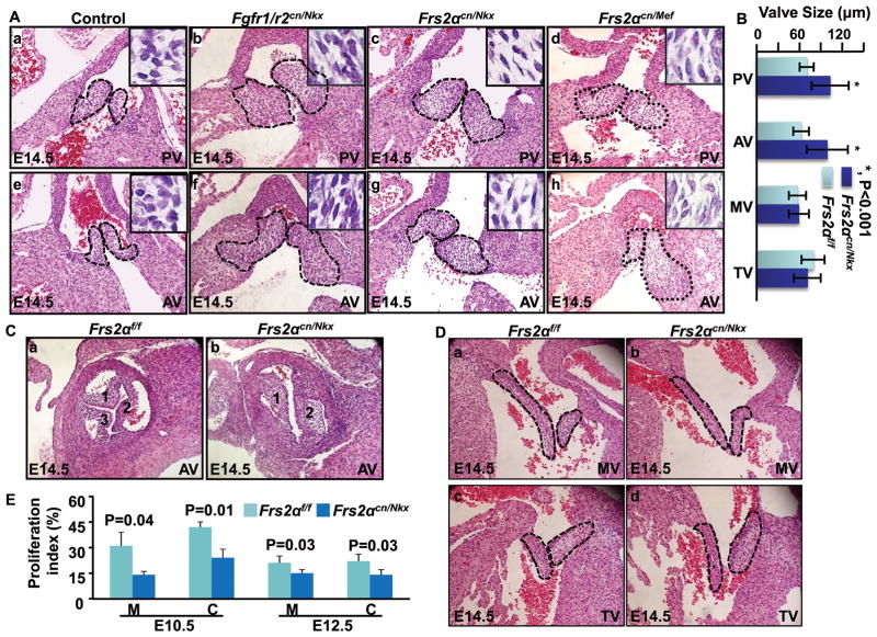
Figure 1. Disruption of the FGF signaling axis leads to OFT valve hyperplasia.
A, Transverse sections of E14.5 embryos were H&E stained to demonstrate enlarged OFT valves. Pulmonary and aortic valves are highlighted with dotted lines. Inserts are enlarged pictures of the valve. B, The embryos were serially sectioned, and the valve dimensions were measured in every five sections. Averages of three largest values were calculated from each embryo. Data represent an average dimension of at least 10 valves and are expressed as means ± standard deviation. C, Coronal sections of embryos were H&E stained demonstrating bicuspid aortic valves in E14.5 Frs2αcn/Nkx embryos. D, Transverse sections of E14.5 embryos were H&E stained demonstrating atrioventricular valves. E, BrdU incorporation demonstrating compromised cell proliferation in Frs2αcn/Nkx OFTs. Percentages of positive cells in the OFT myocardium and cushions from four embryos were calculated and expressed as means ± standard deviations. Fgfr1/r2f/f, Fgfr1 and Fgfr2 double floxed embryos; Fgfr1/r2cnNkx, Fgfr1/Fgfr2 double conditional ablation with Nkx2.5Cre; Frs2αflox, Frs2α floxed, Frs2αcn/Nkx, Frs2α conditional ablation with Nkx2.5Cre; Frs2αcn/Mef, Frs2α conditional ablation with Mef2CCre; PV, pulmonary valve; AV, aortic valve; MV, mitral valve; TV, tricuspid valve; C, cushion; M, myocardium.

