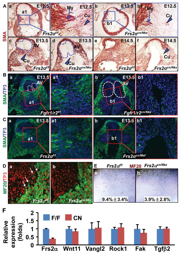
Figure 3. Ablation of the FGF signaling axis leads to defective myocardialization and SM differentiation in OFT cushions.
A-C, Transverse sections of embryos were immunostained with anti-SMA antibody demonstrating defects in myocardialization and SM differentiation. The specifically bound antibodies were visualized either with peroxidase activities (A) or fluorescent dyes (B&C). Arrows indicate migrating myocardial cells (My), and arrow heads indicate cushion mesenchymal cells (Cu) undergoing SM differentiation. Dotted lines outline the valve primordia. To-Pro3 (TP3) was used for nuclear counterstaining. D, Immunostaining with MF20 antibody revealed that the myocardial cells in control OFT were elongated and had long lamellipodia extending into the cushion mesenchyme as indicated by arrows (a); reduced lamellipodia were observed in Frs2αcn/Nkx OFT myocardial cells (b). E, Myocardial cells isolated from E13.5 OFT migrating through the membranes were identified by immunostaining with MF20 antibody (c–d). The ratios of migrated MF-20 positive cells over total MF-20 positive cells were calculated from 3 individual samples and were expressed as means ± standard deviations (e). F, Real-time RT-PCR analyses of E12.5 OFTs. Data are normalized to GAPDH and expressed as folds of changes over wide type samples.

