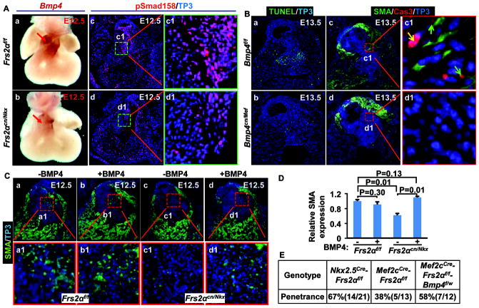
Figure 6. FRS2α-mediated signals regulate OFT cushion remodeling via BMP4.
A, Whole-mount in-situ hybridization with E12.5 embryos demonstrates reduced Bmp4 expression in Frs2αcn/Nkx OFT (panels a and b). Red arrows indicate Bmp4 expression. Panels c and d, transverse sections of E12.5 embryos were immunostained with antibody against phosphorylated Smad1/5/8. B, Apoptotic cells in transverse sections of E13.5 embryos were detected with TUNEL assays (a, b). Panels c and d, double immunostaining of transverse sections of E13.5 embryos with anti-SMA and anti-caspase-3 antibodies. Green arrows indicate cells with moderate caspase-3 activation and SMA expressions, and yellow arrow indicates strong caspase-3 activation. C, Embryonic hearts collected at E12.5 were cultured overnight in the presence or absence of 10 ng/ml of BMP4. Paraffin sections of the hearts were then immunostained with anti-SMA antibodies. D, Real-time RT-PCR analyses of SMA expression in ex vivo cultured OFTs. Data were normalized to GAPDH and expressed as folds of changes over untreated samples. E, Ablation of one Bmp4 allele increases the penetrance of OFT valve defects in Frs2αcn/Mef embryos.

