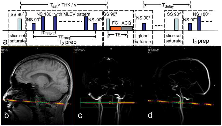
Figure 1.
(a) Pulse sequence diagram for measuring blood T2 in the internal jugular vein (IJV). There are 4 blocks within each TR: slice-selective (SS) saturation, non-selective (NS) T2 prep, SS excitation/acquisition, and NS saturation. Tsat = 200 ms, τCPMG = 10 ms, TEprep = [20, 40, 80, 160] ms, and TE = 15 ms, Tdelay = 2 s. (b–c) Localization of IJV: (b) Anatomical scout image with sagittal view. Angiographic scout images with coronal view (c) and with sagittal view (d) are also shown. The orange line indicates the center of the imaging slice (red) perpendicular to the neck vessels in both coronal view and sagittal view. The green box is the localized shimming box that aligns with the imaging slice and is centered at the side of the largest IJV of the subjects.

