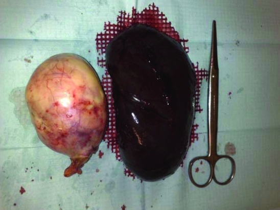
Figure 2.
Macroscopic aspect of the right anexial lesion and spleen after surgical removal. The ovary is transformed into a uniloci cystic formation with 140×90×80 mm, fine wall, pink and smooth external surface and tarnished internal surface with pasty yellow material with hair adherent. Spleen with complete capsule and marked parenchyma congestion.

