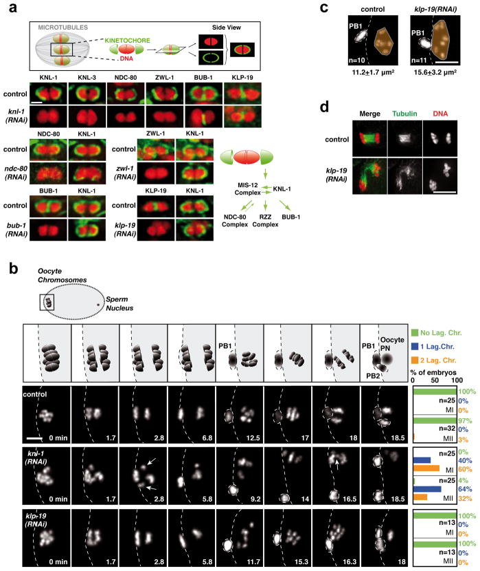
Figure 1. Cup-shaped meiotic kinetochores are assembled by a KNL-1-dependent mechanism and are required for accurate meiotic chromosome segregation.
(a) Top: Schematic of cup-shaped meiotic kinetochores. Bottom. Localization of 6 kinetochore components and the chromokinesin KLP-19 on individual meiosis I bivalent chromosomes (see also Fig. S1). Schematic summarizes assembly dependencies for the meiotic kinetochore. Scale bar, 1 μm. (b) Top row: Schematic of chromosome segregation during meiosis I and II. (PB1: First Polar Body; PB2: Second Polar Body; PN: Pronucleus). 3 Bottom rows: stills from movies of fertilized oocytes expressing GFP-histone-H2b. Time relative to Metaphase I is in the lower right corner of each panel. White arrows indicate lagging chromosomes during anaphase I and II in the KNL-1 depletion. Quantification of lagging chromosomes during anaphase I (MI) and anaphase II (MII) is on the right. Scale bar, 5 μm. (c) KLP-19 depletion causes increased chromosome dispersal in late anaphase that is correlated with spindle instability at this stage. (d) Area occupied by the chromosomes (orange) was measured at a similar time after anaphase I onset (Control, 11.4±1.2 min; KLP-19-depleted, 11.9±0.6 min); spindle instability was observed in 8/8 KLP-19-depleted fixed oocytes at this specific cell cycle stage. Scale bars, 5 μm.

