
Fig. 1. Mice with global deletion of the CCM3 gene (CCM3-KO) die at E8.5 and display defects in VEGFR2 signaling, vasculogenesis and hematopoiesis.
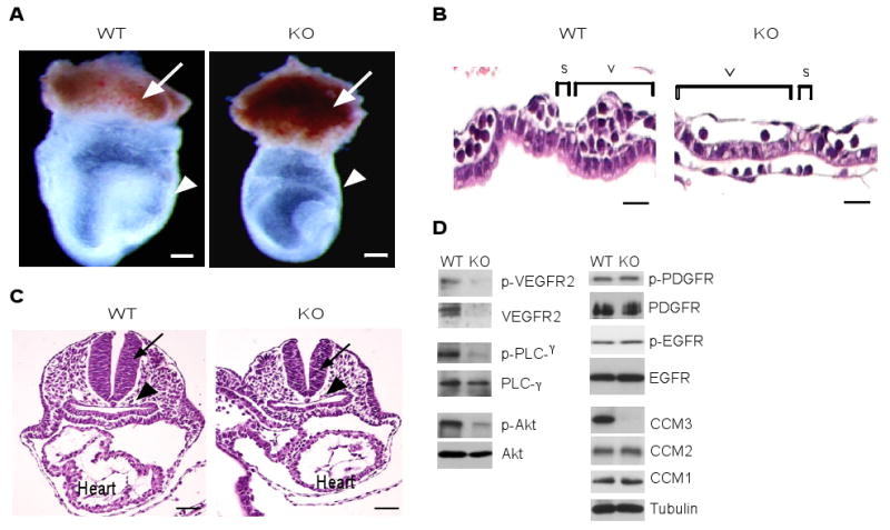
(A) Appearance of wild type and CCM3-KO embryos at E8.0. Embryos were freshly dissected, then photographed. The ectoplacental cone, yolk sac and embryo (inside) are indicated. Scale bar, 200 μm. (B) H&E stained cross-sections of the yolk sac. Yolk sacs from WT and CCM3-KO mice were embedded, followed by H&E staining. The cross-sectional length of the yolk sac vessel, adjacent and parallel to the endoderm layer (v), and the empty space between the vessels (s) are indicated. Scale bar, 20 μm. (C) H&E staining of embryos. Neural tube and dorsal aorta are indicated by an arrow and an arrowhead, respectively. The heart is also labeled. Scale bar, 100 μm. (D) Defects of VEGFR2-dependent signaling in CCM3-KO embryos. Whole embryos of wild type and CCM3-KO were subjected to Western blotting with various antibodies against VEGFR2 signaling molecules, PDGFR, EGFR, and the CCM proteins, as indicated. 5 other embryos of wild type or KO gave similar results. The relative abundance of VEGFR2, p-VEGFR2, the ratios of p-PLC-γ/PLC-γ and p-Akt/Akt are shown, with WT group as 1.0. Data are mean ± SEM (n=6). *, p<0.05.