
Fig. 2. Mice with a vascular endothelial deletion of the CCM3 gene (CCM3-ecKO) display defects in angiogenesis and hematopoiesis.
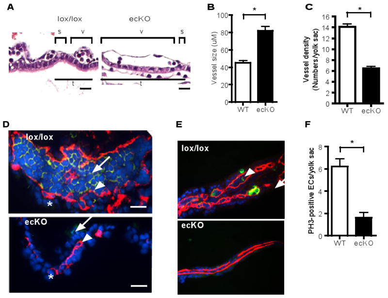
(A to C). H&E stained cross-sections of the yolk sac at E8.5. Yolk sacs from CCM3lox/lox and CCM3-ecKO mice were embedded, followed by H&E staining. The cross-sectional length of the yolk sac vessel, adjacent and parallel to the endoderm layer (v) and the empty space between the vessels (s) as well as total vessel length (t) are measured. The vessel size (μM) and vessel density (number of vessels/yolk sac) are quantified. Scale bar, 20 μm. (D). CCM3-ecKO yolk sacs at E8.5 exhibit reduced VEGFR2 staining. Vessel endothelial cells exhibit positive staining for VEGFR2 (red), whereas blood cells exhibit positive staining for Ter-119 (green). Nuclei were counterstained by DAPI (blue). Scale bar, 10 μm. (E-F) CCM3-ecKO yolk sacs at E8.5 exhibit reduced proliferation. Yolk sacs were stained for phospho-histone-3 (PH3, green) and endothelial cell marker CD31 (red). Blue: DAPI. Scale bar, 100 μm. PH3-positive endothelial cells are indicated by an arrow, with quantification shown in F. PH3-positive blood cell by an arrowhead. Data are mean ± SEM (n=5). *, p<0.01.