
Fig. 3. A vascular endothelial deletion of the CCM3 gene disrupts vascular integrity in mice.
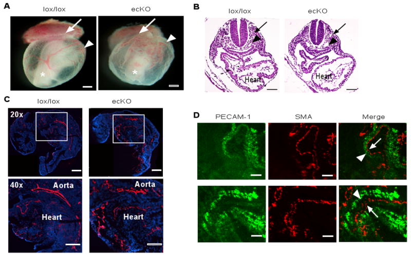
(A) Appearance of WT and CCM3-ecKO embryos at E9.0. Embryos were freshly dissected and photographed. The ectoplacental cone, yolk sac and embryo (inside) are indicated. Scale bar, 200 μm. (B) Histological analysis (H&E staining) of embryos at E9.0. The dorsal aorta, cardinal vein and heart are indicated. Scale bar, 100 μm. (C) Immunostaining (CD31, red) of embryos at E9.0. Blue: DAPI. The aorta and heart are indicated. Scale bar, 200 μm. (D) Co-staining of embryos with antibody against CD31 and SMA. Dissociation of the endocardium (CD31-positive) from the myocardium (SMA-positive) in the hearts of CCM3-ecKO mice at E9.0 is indicated by an arrow. 5 other embryos of wild type or KO gave similar results. Scale bar, 50 μm.