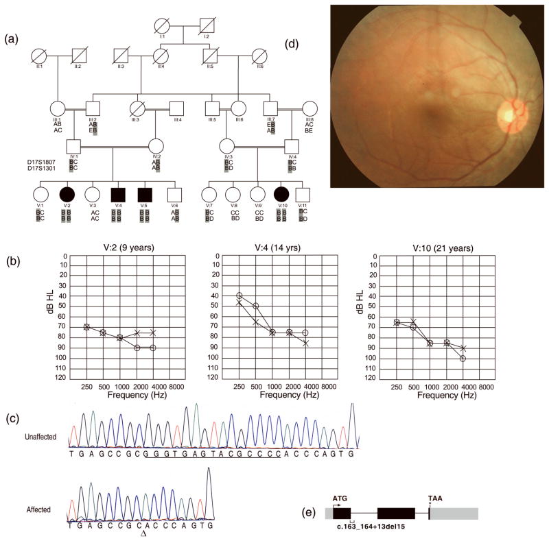
Figure 1.
(a) Pedigree of family HLRB12. Haplotypes for two closest markers to SANS are shown for the genotyped members. Alleles of the two markers were homozygous for all affected individuals. The ancestral chromosome with the SANS mutation is shaded in gray (b) Audiograms of three affected individuals of family HLRB12 indicate moderate to severe hearing loss. Age at the time of audiometry is indicated on tops of the audiograms. “o” indicates air conduction for right ear, while “x” indicates air conduction for left ear (c) A fifteen nucleotide deletion was observed in SANS at the first exon-intron junction in DNA of affected individuals. “Δ” indicates the start of deletion. Deleted bases are underlined in the trace from a control. (d) A representative funduscopic image from a patient with atypical Usher syndrome in family HLRB12. No bone spicules were observed (e) Diagrammatic depiction of SANS. Black boxes represent translated exons while gray boxes denote untranslated parts of the exons. The two introns are represented by horizontal lines. The position of the deletion of 15 nucleotides identified in family HLRB12 is shown by a bracket under the depicted exon 1 and the following intron. The arrow and the asterisk denote the start and stop codons in SANS, respectively.

