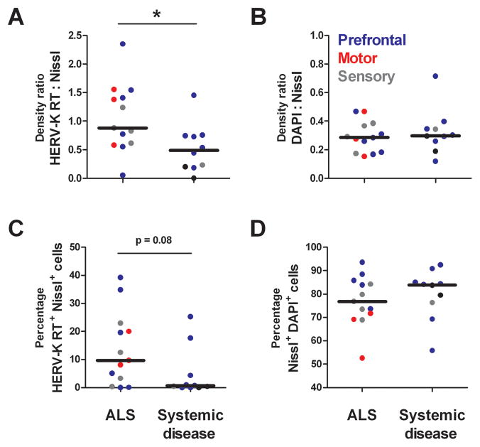
Figure 5. Quanitative analysis of HERV-K RT immunostaining in cortical brain tissue of patients with ALS or systemic disease.
Patients with ALS exhibit significantly more HERV-K RT protein expression, as measured by staining density, than cortical tissue from patients with systemic disease (Panel A). The same trend was evident when cell counting was used to quantify HERV-K RT expression (Panel C). As an experimental control, the Nissl to DAPI ratios demonstrate that there is similar neuronal staining in both ALS and patients with systemic disease (Panels B and D). Asterisk represents p<0.05.

