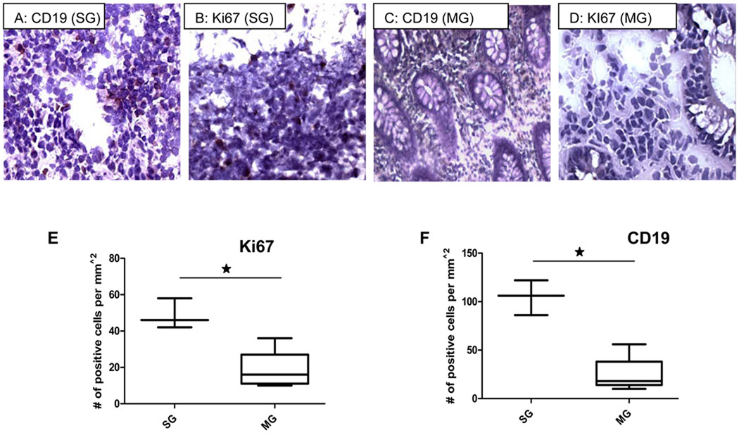
Figure 4.
Higher levels of CD19+ cells (B-lymphocytes) (A,C,F) and Ki67 (B,D, E) were seen in highly inflamed bowel in SG patients compared to biopsy samples from MG patients. Lower levels of CD19 positive B-lymphocytes were observed in biopsy samples from patients responding to therapy (MG) with CD (4C). No staining was observed in the tissue sections not treated with primary antibody. (* indicates p<.05 using the unpaired t-test; SG indicates patients requiring surgical management; MG indicates patients responding to medical therapy).

