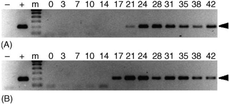
Fig. 3.
Transstadial transmission of E. canis to dogs by male R. sanguineus. PCR assays were performed on peripheral blood from dogs AHK (panel A) and AIP (panel B). Lanes containing the 200 bp target amplicon (arrowhead) were considered PCR-positive. For each panel, template-free reactions served as negative controls (-), template DNA (1 ng) collected from E. canis-infected DH82 cells served as positive control (+) and 0-42 indicate days post-tick attachment. The molecular size standard (m) is a 100 bp ladder.

