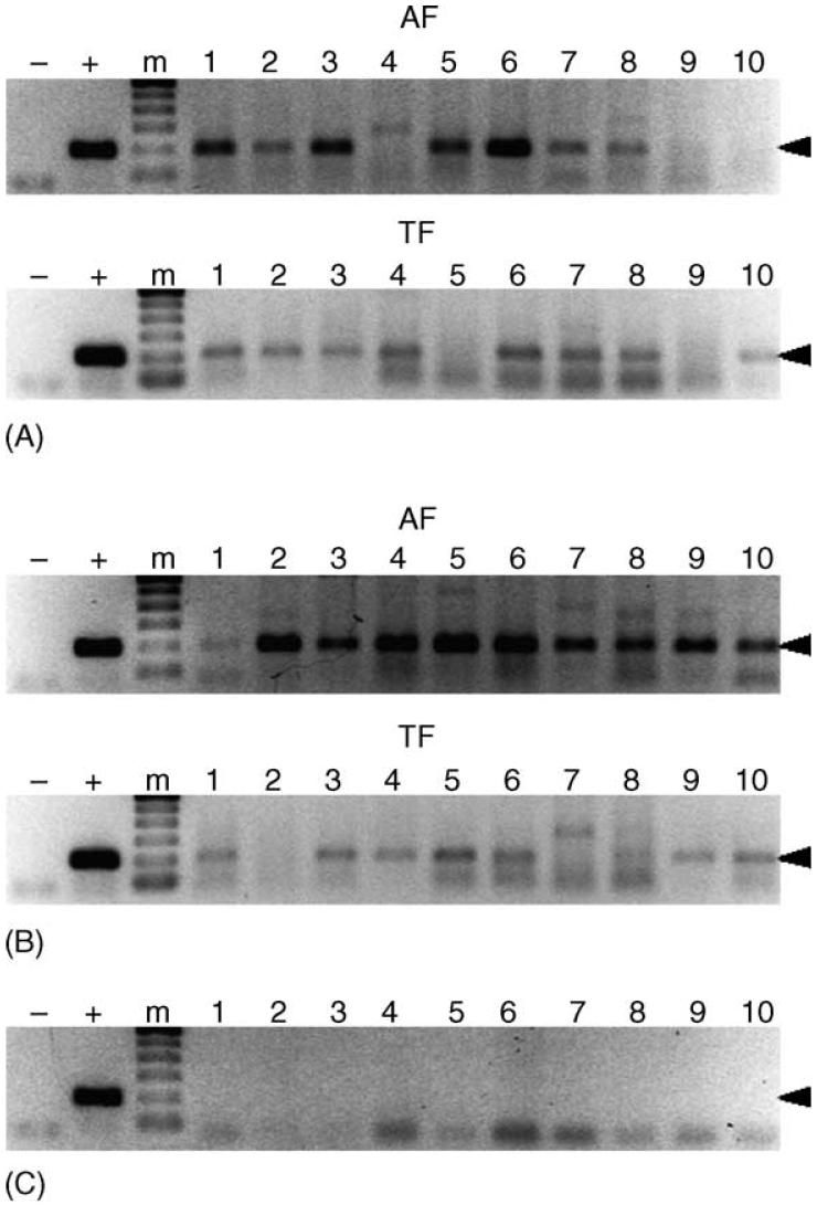
Fig. 4.
Detection of E. canis in intrastadially exposed male R. sanguineus. Panels A and B represent male R. sanguineus exposed to E. canis on dogs ATK and BAA, respectively, which were assayed after acquisition (AF) and transmission (TF) feeding on dogs AHG and AUF, respectively. Panel C represents male R. sanguineus allowed feed on the negative control, dog AXM. Samples resulting in a 200 bp amplicon (arrowhead) were considered PCR-positive. For each panel, template-free reactions served as negative controls (-), template DNA (1 ng) collected from E. canis-infected DH82 cells served as positive control (+), and lanes labeled 1-10 represent individual ticks. The molecular size standard (m) is a 100 bp ladder.

