
Figure 1.
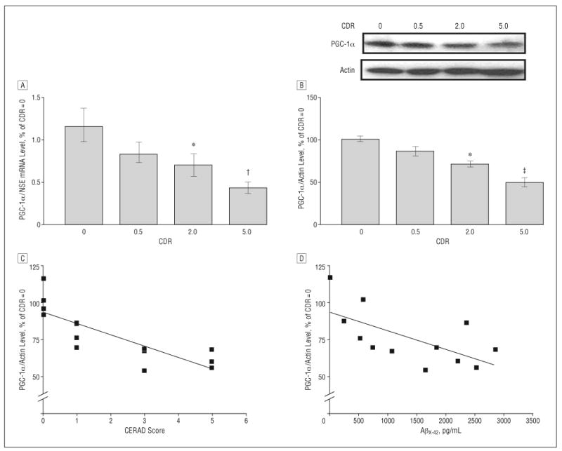
Hippocampal peroxisome proliferator–activated receptor γ coactivator 1α (PGC-1α) expression in the Alzheimer disease (AD) brain decreases as a function of AD dementia and AD β-amyloid (Aβ) neuropathology. A, PGC-1α messenger RNA (mRNA) content in the hippocampal formation (quantified by real-time reverse transcriptase–polymerase chain reaction and normalized by neuron-specific enolase [NSE]) as a function of Clinical Dementia Rating (CDR) representing cognitive normalcy (CDR=0), questionable dementia (CDR=0.5), mild dementia (CDR=2), and severe dementia (CDR=5). B, Western blot confirmation of decreased PGC-1α protein content in the hippocampal formation of AD cases. In A and B, data represent mean (SEM) and are shown as a percentage relative to the CDR=0 group. *P<.05, †P<.003, and ‡P<.01 vs control group by t test. C and D, PGC-1α mRNA expression as a function of Aβ neuritic plaque neuropathology in accord with the Consortium to Establish a Registry for Alzheimer's Disease (CERAD) 4-point scale (n=4, 4, 3, and 3 for CERAD scores of 0, 1, 3, and 5, respectively) for AD (C) or content of Aβx-42 (n=12) (D). Straight line represents best linear regression fit.