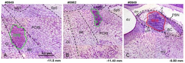
Fig. 1.
Light photomicrograph examples of retrograde tracer injections into the RF and PBN counterstained with cresyl violet (5x). A) Injection of GFB into the IRt. B) Injection of GFB into the PCRt. C) Injection of RFB into the PBN of the same animal depicted in A. The extent of the tracer material is outlined with a green (GFB in A & B) or red line (RFB in C). Central medial (cm), dorsal medial (dm), ventral medial (vm) and ventral lateral (vl) subdivision of the PBN are indicated in this figure. The approximate levels relative to bregma are indicated below each photomicrograph (Paxinos and Watson, 1982). 4V, 4th ventricle; BC, brachium conjunctiva; Gi, gigantocellular reticular formation; IRt, intermediate zone of medullary RF; mV, motor trigeminal nucleus; NST, nucleus of the solitary tract; PCRt, parvocellular zone of medullary RF; Sp5, spinal trigeminal tract.

