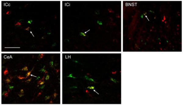
Fig. 5.

High power (60x) photomicrographs of RF (GFB injection) and PBN (RFB injection) projection neurons in the contralateral insular cortex (ICc), ipsilateral insular cortex (ICi), bed nucleus of the stria terminalis (BNST), central nucleus of the amygdala (CeA), and lateral hypothalamus (LH). Arrows show an example of a cell in each forebrain area that projected both to RF and PBN. The scale bar in left top bar equals 50 um.
