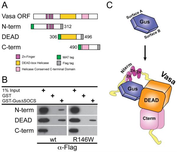
Figure 4. GusΔSOCS-Vasa interacts with discrete segments of Vasa protein.
(A) A graphic representation of each Vasa construct containing an N-terminal Metal Affinity Tag (MAT), C-terminal Flag epitope, corresponding domains and DEAD-box sequence motifs. (B) Gustavus binding domains in Vasa. Bacterial extracts containing N-term, DEAD and C-term recombinant Vasa proteins were incubated with immobilized GST, GST-GusΔSOCS wt or GST-GusΔSOCS R146W and bound proteins were analyzed by immunoblotting with anti-Flag antibodies. (C) A bipartite Gustavus-Vasa binding model in S. purpuratus. The gustavus B30.2/SPRY domain is shown in blue with binding surface A and binding surface B indicated as predicted previously (Woo et al., 2006a). A schematic representation of the S. purpuratus vasa DEAD box domain in orange, C-terminus domain in pink, N-terminus CCHC zinc knuckles in purple and unstructured glycine-rich flexible sequence in yellow.

