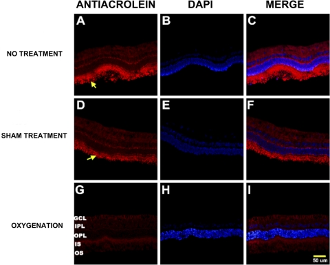
Figure 6.
Photomicrographs of vertical retinal sections from standardized regions (4 disc diameters below the optic disc) in the retina processed for anti–acrolein immunoreactivity (A, D, G) with the nuclear counterstain DAPI shown in images (B, E, H) and merged images (C, F, I).The secondary antibody was Texas Red-conjugated anti-mouse IgG. High expression of the acrolein was shown in both the no treatment (A) and the sham treatment (D) groups compared with the oxygenation group (G). GCL, ganglion cell layer; IPL, inner plexiform layer; ONL, outer nuclear layer; IS, inner segments of the photoreceptor layer; OS, outer segments of the photoreceptor layer. Arrows: areas of increased acrolein expression in the photoreceptor IS and OS.

