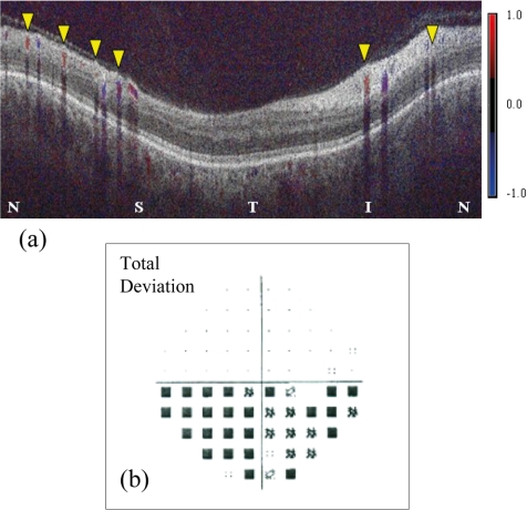
Figure 4.
Color Doppler OCT of an eye with NAION. (a) Superior retinal hemisphere (left half of image) showed lower flow, 13.6 μL/min in 4 veins (yellow arrowheads), compared with the inferior retinal hemisphere (right half of image), where the flow was 18.7 μL/min in two veins (yellow arrowheads). N, nasal; S, superior; T, temporal; I, inferior. Visual field total deviation map (b) showed altitudinal defect in the inferior field (corresponding to the superior retinal hemisphere).

